Table of Contents
Sense of Sight Introduction
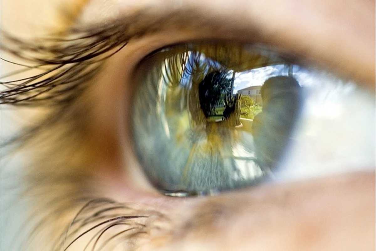
The sense of sight is unique to the five senses that the human being has and, perhaps, one of the most important. When we say that “an image is worth a thousand words”, it is because we live in a world where what we can see, interpret and identify in our environment is fundamental for us.
This visual information that we capture thanks to the sense of sight is because the eye is one of the most evolved organs that the human body has.
The Eye Receptor Organ
The eye is an organ situated in the skull’s bony cavity. Its inside part is formed with eyelids and eyelashes, which protect it from entering any substances and keep it clean, lubricant, and moist.
Parts of the Eye
The visual system detects light stimuli (electromagnetic waves), distinguishing between two characteristics of light, its intensity and wavelength (colors). However, the morning, before reaching the retina, passes through the different parts of the eye: the cornea, the aqueous humor, the pupil, the crystalline or natural lens of the eye and the vitreous humor.
In addition, the retina contains two types of photoreceptor cells. The so-called rods (responsible for peripheral and night vision) and cones (are sensitive to the color of light).
How are Images Formed?
While light passes through the cornea and lens, an inverted, actual image forms on the retina through the pupil, this inversion occurs due to different densities of the areas that light passes through so that the upper light rays are project on the lower part of the retina and the lower ones on the upper part.
According to most theories, the optic nerve carries this stimulus to the cerebral cortex, where the interpretation of the message is made through a psychic-chemical process.
Eye Anatomy
Sclerotic
Sclera, or the white colour of our eyes, is a membrane formed by collagen that, in addition to protecting the eye, regulates the passage of light. This part of the eye has muscles that help us move the eyeballs, and the front is covered with the cornea.
Cornea
The cornea is a transparent layer of avascular tissue of the eye that contains five layers: the first epithelium, Bowman’s membrane, the stroma, Descemet’s membrane, and the endothelium.
Its two main functions are protecting the intraocular content and the refraction of light. It represents almost 80% of the total refractive power.
Coroides
The choroid is a dark membrane between the sclera and the retina. Its primary mission is to nourish the retina through its numerous blood vessels.
Ciliary Body
The ciliary body consists of a circle of tissue surrounded by a natural eye lens or crystalline lens. The muscle fibers do the maintenance of the shape of the eye. It is also responsible for secreting aqueous humour in the anterior eye segment—the pupil’s magnitude and the lens’s form change when the eye focuses on an object.
Pupils
The pupil is the part of the eye, or black dot (hole) that we have in the iris, which contracts (miosis) and dilates ( mydriasis ) to regulate the passage of light that will finally reach the retina. In the dark, the pupil dilates to capture more light and, the opposite, when the environment is very bright.
Iris
The iris is the cultured circle around the pupil that allows the pupil to dilate. This eye has color thanks to cells with a pigment called melanin and melanocytes.
Retina
The retina, for its part, is in charge of receiving light stimuli through its receptor cells: rods (light intensity) and cones (colour). The fovea is the one that contains the cones, which is where the light beam from the visual axis arrives.
The retina’s role is fundamental for the sense of our sight since it will depend on how that image reaches the brain, interprets it and becomes the vision that we are going to see later.
Aqueous Humor
The aqueous humor is a transparent fluid between the lens and the cornea. The function of the aqueous humor is to maintain the shape of the cornea by adding pressure to it and keeping the curved shape outside.
Crystalline or Lens
The lens is the natural lens that our eye has and that overtime loses elasticity and becomes cloudy, forming a cataract. It is in charge of regulating the focus, allowing greater or lesser sharpness, adapting its shape from more concave to more convex thanks to the ciliary muscles.
Vitreous Humor
The vitreous humour is the jelly-like fluid found in the most eyeball. It maintains its round shape between the retina and the back of the lens.
Optic Nerve
The optic nerve is responsible for transferring signals and information from the eye to our brain to be processed by the visual cortex, the hypothalamus and the occipital lobe.
Primary Eye Care
The sense of sight and, therefore, the state of our eyes is essential for the performance of our tasks daily. However, being such a delicate and small organ, it is vitally important to give it the necessary care. At least a complete ophthalmological examination is require every year to rule out any pathology that may be associate with them.
But in addition, some of the main recommendations to follow are:
- Take care of your diet: through a diet rich in vitamins A and C, essential for sight, such as carrots, asparagus, apricots and nectarines, as well as dairy products.
- Keep our eyes hydrated: mainly if we are use to working for many hours in front of the computer or very dry or humid environments.
- Correct lighting: visual fatigue can be prevent with adequate lighting. This way, we avoid excessive effort when working or reading, especially in office environments.
- Wear sunglasses with filters for ultraviolet rays every month of the year.
- Learn to relax your eyes: specialists recommend focus change techniques.
- And, of course, go to the ophthalmologist regularly to prevent any problem.